sponsored content
September 1, 2023
By Debbie Jones, FCOptom, FAAO, FCBLA
Axial length measurement should be considered an essential part of the assessment of patients at risk of developing myopia, as well as those who are already myopic.

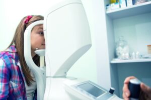
Photo Credit: bluecinema, Getty Images
As eye care professionals embrace the need to manage myopia with more than just a simple spherical optical correction, it is important to understand that the examination of patients involves more than just an assessment of refractive error. The measurement of axial length plays an important role in the decision-making process when managing our young myopic and pre-myopic patients.
Axial length (AL) is measured in millimeters from the anterior cornea to the retinal pigment epithelium and is the most significant contributor to refractive error and potential myopia-related visual impairment.1
There are two basic methods to measure AL, ultrasound and optical biometry.2 The use of A-scan ultrasound, which requires contact with the cornea, has been largely superseded by modern instrumentation that avoids contact and has improved accuracy and ease of use. There are a variety of instruments available to measure AL, broadly divided into stand-alone instruments and multifunction instruments. Multifunction instruments have the advantage of occupying a small footprint, while offering the option of AL measurement along with other clinical measures/assessments, such as autorefraction, topography, support for contact lens fitting, and dry eye assessment. In general, optical biometers are more reliable than ultrasound biometers as they produce more repeatable measurements compared to ultrasound biometers. For instance, a study reported a smaller test-retest difference for the IOLMaster optical biometer compared to A-scan (0.004mm versus 0.042mm).3
The AL of a newborn eye is approximately 16.8mm, with an emmetropic adult eye being 23.8mm.4,5 There is, therefore, natural growth to be expected during childhood. The typical changes in AL are 0.15mm to 0.20mm/year up to age 8, and then 0.06 to 0.15mm/year through to the early teen years.6,7,8 Progression outside of this rate should give cause for concern, as the peak rate of axial elongation may occur two to four years prior to evidence of a myopic refractive error.8 An AL of 23.07mm in a 6-7 year old and a change of 0.74mm across a three-year time period are outside the norm and suggestive of future myopia development.9
Identifying those patients at risk of developing myopia by careful monitoring of axial length can result in discussions around the need for early intervention with simple lifestyle modifications, such as spending more time outside, in an attempt to delay the onset of myopia.10 It has been shown that those patients with a myopic refractive error prior to the age of 10 are at a greater risk of developing high myopia in their adult years, and therefore delaying the onset of myopia is hugely beneficial.11 The severity of myopia-related visual impairment increases with increasing axial length.1 For example, patients with an axial length ≥ 30mm have a 90 times greater chance of developing uncorrectable visual impairment compared to an individual with an emmetropic axial length.1
There are large data sets of normative axial length available,6,12 providing an opportunity to compare the patient in the chair with a matched data set (of both gender and ethnicity). This gives an indication of possible trajectory and risk of progression. Comparison to normative data is invaluable when discussing the need for myopia management with parents in being able to demonstrate the potential refractive error outcome with no myopia management intervention.6,12,13 The clinical application of myopia management can be simple to explain in terms that are easily understood by parents, relating to other health metrics. For example, children below the 50th percentile for AL for age are less likely to become myopic than those over the 50th percentile. Many of the multifunction instruments designed to measure AL have normative data sets included in their software, facilitating comparison of the patient being examined against matched norms, and they can also be used to monitor progression. The ability to print out AL reports for parents can improve their confidence in the myopia management process.
As myopia management continues to be recognized as an important part of primary eye care, we can anticipate the availability of an increased number of myopia management products on the market. Therefore, monitoring AL is an essential part of assessing the success of the chosen myopia management modality. In the case of orthokeratology, it is the only effective way to assess efficacy of the treatment for a patient.14 Even for other modalities, it is imperative that the effect of the treatment on AL is as meaningful as it is for refractive error. Consistent with this, both AL and refractive error changes are reported primary outcome measures in clinical studies of new and existing myopia management modalities.15
There is no doubt that there is a benefit to undertaking myopia management, and measurement of AL is a major factor in managing myopia. Axial length measurement should be considered an essential part of the assessment of patients at risk of developing myopia, as well as those who are already myopic. ECPs must understand that myopia is more than just a myopic refractive error.
To read more about the importance of axial length measurement and CARE (Cumulative Absolute Reduction in axial Elongation), please see Johnson & Johnson Vision’s previous article by Dr. Noel Brennan.
 |
Debbie Jones, FCOptom, FAAO, FBCLA, is a Clinical Professor at the School of Optometry and Vision Science and a Clinical Scientist at the Centre for Ocular Research & Education (CORE) at the University of Waterloo. Her main area of clinical focus is in pediatric optometry and her main area of research activity is in the area of myopia control. Debbie has a keen interest in the eye care needs of indigenous peoples and has provided eye care to a remote community in Northern Ontario on a number of occasions. Trained in the U.K., Debbie has been a faculty member at the School of Optometry and Vision Science for 25 years. She is formally a partner in an award winning private practice in the U.K., has published articles in optometric journals, and regularly presents at optometric conferences worldwide. She is a Fellow of the British College of Optometrists, the British Contact Lens Association, and the American Academy of Optometry. |
References
- Tideman, J.W., et al., Association of Axial Length With Risk of Uncorrectable Visual Impairment for Europeans With Myopia. JAMA Ophthalmol, 2016. 134(12): p. 1355-1363.
- Chia TMT, Nguyen MT, Jung HC. Comparison of optical biometry versus ultrasound biometry in cases with borderline signal-to-noise ratio. Clin Ophthalmol. 2018 Sep 10;12:1757-1762. doi: 10.2147/OPTH.S170301. PMID: 30237695; PMCID: PMC6136410.
- Hussin, H.M., et al., Reliability and validity of the partial coherence interferometry for measurement of ocular axial length in children. Eye (Lond), 2006. 20(9): p. 1021-4.
- Gordon, R.A. and P.B. Donzis, Refractive development of the human eye. Arch Ophthalmol, 1985. 103(6): p. 785-9.
- Atchison, D.A., et al., Eye shape in emmetropia and myopia. Invest Ophthalmol Vis Sci, 2004. 45(10): p. 3380-6.
- Tideman, J.W.L., et al., Axial length growth and the risk of developing myopia in European children. Acta Ophthalmol, 2018. 96(3): p. 301-309.
- Zadnik, K., et al., Normal eye growth in emmetropic schoolchildren. Optom Vis Sci, 2004. 81(11): p. 819-28.
- Mutti, D.O., et al., Refractive error, axial length, and relative peripheral refractive error before and after the onset of myopia. Invest Ophthalmol Vis Sci, 2007. 48(6): p. 2510-9.
- McCullough, S., et al., Axial growth and refractive change in white European children and young adults: predictive factors for myopia. Sci Rep, 2020. 10(1): p. 15189.
- Xiong, S., et al., Time spent in outdoor activities in relation to myopia prevention and control: a meta-analysis and systematic review. Acta Ophthalmol, 2017. 95(6): p. 551-566.
- Hu, Y., et al., Association of Age at Myopia Onset With Risk of High Myopia in Adulthood in a 12-Year Follow-up of a Chinese Cohort. JAMA Ophthalmol, 2020. 138(11): p. 1129-1134.
- He, X., et al., Normative data and percentile curves for axial length and axial length/corneal curvature in Chinese children and adolescents aged 4-18 years. Br J Ophthalmol, 2021.
- Sanz Diez, P., et al., Growth curves of myopia-related parameters to clinically monitor the refractive development in Chinese schoolchildren. Graefes Arch Clin Exp Ophthalmol, 2019. 257(5): p. 1045-1053.
- Vincent SJ, Cho P, Chan KY, Fadel D, Ghorbani-Mojarrad N, González-Méijome JM, Johnson L, Kang P, Michaud L, Simard P, Jones L. CLEAR – Orthokeratology. Cont Lens Anterior Eye. 2021 Apr;44(2):240-269.
- Wolffsohn, J.S., et al., IMI – Clinical Myopia Control Trials and Instrumentation Report. Invest Ophthalmol Vis Sci, 2019. 60(3): p. M132-M160.
PP2023OTH5422


