October 2, 2019
By Pavan K. Verkicharla, BS Opt., PhD
Scientist, Myopia Research Lab – L V Prasad Eye Institute, India
Visiting Scientist – BHVI, Australia
I will start by quoting Holden et al. (2015): “Optometry should be on the frontline of this (myopia) battle. It not only has a responsibility to provide the best care to patients but to remain informed about epidemiological trends and understand the latest range of treatment options for progressive myopia.”
As optometrists, we are the most responsible individuals of the eye care profession for the management of myopia, and thus any myope should undoubtedly be managed in any regular primary care optometric practice.
All it needs is to consider 4Ms as a mantra for successful myopia management in any optometry practice: Master – Measure – Monitor – Manage.
- Be a MASTER. We need to first educate ourselves about the variants of myopia and its management. Understand that myopia has multi-factorial a Myopigenesis is a result of the interplay between various factors related to genetics and environment. Therefore, each myope is different and may present to the clinic with something different from their history, either related to genes or the environment.
- Measure/document the ‘X’ factors, i.e., estimate/quantify the possible myopic risk factors.
Documenting/quantifying the risk factors play a key role in deciding on the appropriate anti-myopia (treatment) strategy.
It is vital to have a clinical protocol where all significant myopia related risk factors get documented. The protocol can be divided into three sections, i.e., history, present, and future.
History:
- Myopic parents: Increase the child’s risk of myopia by 1.5 to 7.5 times (based on various studies).
- Environment: Recent literature blames environmental factors for myopia such as reading in dim illumination, limited time outdoors (a study showed that East Asian children spend <30 minutes per day vs. >2 hours by Caucasians in Australia), prolonged near work due to striving for academic excellence or the intense education system (causing stress in the equator of eye due to ciliary muscle contraction) and the advancements in technology which got everything closer to the eyes.
- Age of onset of myopia: The earlier the myopia develops, the greater the risk of high myopia at later age (during adulthood). Thus, careful and close monitoring should be considered in individuals with myopic or emmetropic refractive error at the age < 10 years (see the concept of “pre-myopia” as defined by the International myopia institute).
Present and Future:
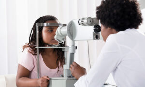
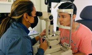
- Central refraction and axial length: The entire myopia management strategy is based on a single metric relating to the change in refractive error and/or axial length over a specific time and best ways to slow it down. Thus, any optometry practice should have a facility to record refraction and axial length. Commercially available non-contact biometers help record axial length accurately in children.
- Peripheral refraction (peripheral hyperopic refraction increases the risk of myopia development and progression).
- Accommodation (is there any lag?) and phoria (is there any esophoria for near?)
- Posterior segment: There are a lot of portable, cost-effective fundus cameras that can be used in regular clinics to capture any retinal or choroidal pathologic myopia lesions.
- Monitor the minor refractive error-related optical and structural changes in the eye that could potentially trigger onset or progression. Although there are no universal guidelines that can be applied to all myopes or “pre-myopes,” it is important to ensure that monitoring or future follow-ups are based on past (history) and present (measurements) and need to be 3-6 monthly during the progression phase. Additionally, in those myopes prescribed with anti-myopia devices/strategies, close monitoring is also necessary to ensure that the device/strategy performs optimally and without any complications.
- Manage myopia with appropriate anti-myopia strategies. The goal of any anti-myopia strategy is to a) prevent myopia or b) delay the onset of myopia or c) stop or slow the progression of myopia. Anti-myopia strategies are broadly classified as optical, environmental/behavioral, and pharmacological interventions. And, their working principles centre on counteracting specific risk factor(s) of myopia. Thus, any strategy to counteract myopia progression in a child should be decided for each child, having given due consideration to the “X”-factors, and not simply based on on-axis refraction alone. Here are some of the optical and environmental options and their efficacy based on evidence.
Bifocals and Progressive Addition Lenses (PALs): Three potential working principles for bifocal spectacle lenses acting to slow myopia are:
- Reduction in lag of accommodation;
- Reduction in accommodative demand thereby reducing ciliary muscle tension and scleral stress; and
- Reduction in peripheral hyperopic defocus at least in superior retina.
Recent RCT involving executive bifocal lens designs (+1.50 D add alone, and +1.50 D add with 3D Base-In prism) significantly reduced myopia progression in children older than 3 years compared with single vision spectacles (-1.25 D [bifocals] versus -2.06 D [SV]).
Progressive lens (PALs) work similarly to bifocals except for that PALs effectively control accommodative lag/accommodative demand at all distances.
Multifocal-like soft contact lens: Evidence indicates that there is a significant slowing of myopia progression (approximately 25% to 72%) with multi-focal soft contact lens interventions compared to standard single vision spectacles or contact lenses. Multifocal-like soft contact lenses may work by counteracting peripheral defocus, inducing simultaneous defocus or the add power may serve to reduce the accommodative demand.
Orthokeratology (OK) lens: OK lenses flatten the central cornea and steepen the mid-peripheral cornea and result in relative peripheral myopic defocus at the retinal plane. OK lenses are shown to be effective (up to 55% to 63%) in controlling axial elongation compared to single vision spectacle correction. The concern is complications; however, ensuring proper care and regular follow-ups will keep the process smooth.
Time outdoors: A significant reduction in the incidence of myopia was found with improved time outdoors. The underlying mechanism is not clear, and various hypotheses were formulated to explain the effect such as dopamine release in the retina, uniform dioptric visual field, exposure to a specific spectrum of light such as blue, vitamin-D and lastly relaxed accommodation (not doing near work or reading). It might also be that outdoors is protective for myopia (either preventing myopia or delaying the onset of myopia) as children are simply not reading/writing or not performing prolonged near activity.
To conclude, all we need are the 4Ms to successfully manage a myope.
The mantra is: Master – Measure – Monitor – Manage

Pavan K. Verkicharla, BS Opt., PhD, Scientist, Myopia Research Lab – L V Prasad Eye Institute, India; Visiting Scientist – BHVI, Australia
References:
- Holden BA, Jong M, Davis S, Wilson D, Fricke T, Resnikoff S. Nearly 1 billion myopes at risk of myopia‐related sight‐threatening conditions by 2050–time to act now. Clinical and Experimental Optometry. 2015; 98(6):491-3.
- Huang J, Wen D, Wang Q, McAlinden C, Flitcroft I, Chen H, Saw SM, Chen H, Bao F, Zhao Y, Hu L. Efficacy comparison of 16 interventions for myopia control in children: a network meta-analysis. 2016; 123(4):697-708.
- Gifford KL, Richdale K, Kang P, Aller TA, Lam CS, Liu YM, Michaud L, Mulder J, Orr JB, Rose KA, Saunders KJ. IMI–Clinical Management Guidelines Report. Investigative Ophthalmology & Visual Science. 2019; 60(3):M184-203.
- Wildsoet CF, Chia A, Cho P, Guggenheim JA, Polling JR, Read S, Sankaridurg P, Saw SM, Trier K, Walline JJ, Wu PC. IMI–Interventions for Controlling Myopia Onset and Progression Report. Investigative Ophthalmology & Visual Science. 2019; 60(3):M106-31.