June 1, 2020
Trang Truong, BOptom, and Thao Ha, MD
Brien Holden Vision Institute
 Myopic maculopathy is the most frequent complication that leads to irreversible visual impairment and blindness in high myopes. Over the years, a number of classification systems were used to classify the signs and severity of the condition but did not adequately cover all the possibilities. More recently, an international photographic classification and grading system was proposed by Ohno Matsui and colleagues. The new tool was applied in this study to assess a large number of highly myopic cases to describe the condition and explore the risk factors for high myopia.
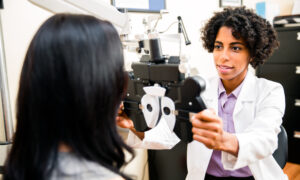
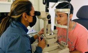
This cohort included 890 myopes with the spherical refraction of −6.00D or worse and was aged 7 to 70 years. The main measurements include the general ocular examination, central axial length, cycloplegic refraction, and color fundus photography following the grades of The International Photographic Classification and Grading System for Myopic Maculopathy. The grading scale consists of five categories following on the severity, including category 0, no myopic retinal lesions; category 1, tessellated fundus only; category 2, diffuse chorioretinal atrophy; category 3, patchy chorioretinal atrophy; category 4, macular atrophy. The clinically significant myopic maculopathy (CSMM) is indicated by category 2 or higher.
Only the right eyes of 884 participants were investigated, while six remaining cases were excluded due to the current retinopathies. After analysis, the distribution of eyes in category 1, category 2, category 3, and category 4 were 20.0 percent (177 eyes), 20.2 percent (178 eyes), 2.6 percent (23 eyes), and 0.2 percent (2 eyes), respectively. Accordingly, CSMM was identified in 203 (22.9 percent) of cases. There was a proportional correlation between the severity of myopic maculopathy and older age, increasing myopia, and longer axial length, but not a correlation with gender. The study also showed that children with early onset of high myopia (7-11 years) have a greater risk of myopic maculopathy development than later-onset patients. Furthermore, it suggests the new cut-off point of pathologic myopia as with spherical equivalent of −8.00D or worse.
The remarkable advantages of this research are the use of a new grading system, a significant sample size and a wide range of age. However, the study was only cross-sectional and was not able to quantify other complications such as posterior staphyloma. Since the study considered only Chinese individuals, it is not entirely clear if the results can be translated to other populations.
Abstract
Xiao O, Guo X, Wang D, Jong M, Lee PY, Chen L, Morgan IG, Sankaridurg P, He M.
Purpose: The purpose of this study was to document the distribution of the severity of myopic maculopathy in a cohort of highly myopic patients and to explore the associated risk factors.
Methods: A total of 890 Chinese high myopes aged between 7 and 70 years (median age 19 years) and with spherical refraction 6.00 diopter (D) or worse in both eyes were investigated. All participants underwent detailed ophthalmic examination. Myopic maculopathy was graded into five categories according to the International Photographic Classification and Grading System using color fundus photographs: category 0, no myopic retinal lesions, category 1, tessellated fundus only; category 2, diffuse chorioretinal atrophy; category 3, patchy chorioretinal atrophy; category 4, macular atrophy. Category 2 or greater were further classified as clinically significant myopic maculopathy (CSMM).
Results: Data from 884 of 890 right eyes were available for analysis. The proportions of category 1, category 2, category 3, and category 4 were 20.0 percent (177 eyes), 20.2 percent (178 eyes), 2.6 percent (23 eyes), and 0.2 percent (2 eyes), respectively. The proportion of CSMM increased with more myopic refraction (odds ratio 1.57; 95 percent confidence interval: 1.46-1.68), longer axial length (odds ratio 2.97; 95 percent confidence interval: 2.50–3.53), and older age (40–70 years compared to 12–18 years, odds ratio 6.77; 95 percent confidence interval: 3.61–12.70). However, there was a higher proportion of CSMM in children aged 7 to 11 years than those aged 12 to 18 years (20.9 percent vs. 11.0 percent, P = 0.008).
Conclusions: Older age, more myopic refraction and longer axial length were associated with more severe myopic maculopathy. Although CSMM was uncommon among younger participants, children with early-onset high myopia have a disproportionately increased risk.
Xiao, O., Guo, X., Wang, D., Jong, M., Lee, P., Chen, L., Morgan, I., Sankaridurg, P. and He, M., 2018. Distribution and Severity of Myopic Maculopathy Among Highly Myopic Eyes. Investigative Opthalmology & Visual Science, 59(12), p.4880.